- (Mon. - Fri 08:00 - 18:00) (Sat 08:00 - 15:00)
- +90 462 227 6100
- [email protected]
A clinical guide to the diagnosis, surgical extraction, and long-term oral health outcomes of impacted wisdom teeth, canines, and premolars.
In ideal oral anatomy, teeth erupt smoothly through the gingival tissue and align naturally within the dental arch, establishing proper masticatory function and aesthetic balance. However, factors such as genetic predisposition, inadequate jawbone space, or structural obstructions from neighboring teeth can prevent a tooth from emerging fully into its correct position. Teeth that remain partially or entirely trapped beneath the gumline or jawbone are clinically classified as impacted. Although this condition is most frequently observed in third molars (commonly known as wisdom teeth), it also regularly affects maxillary canines and premolars. If left undiagnosed during early developmental stages, impacted teeth can cause serious underlying medical complications within the maxillofacial structure.
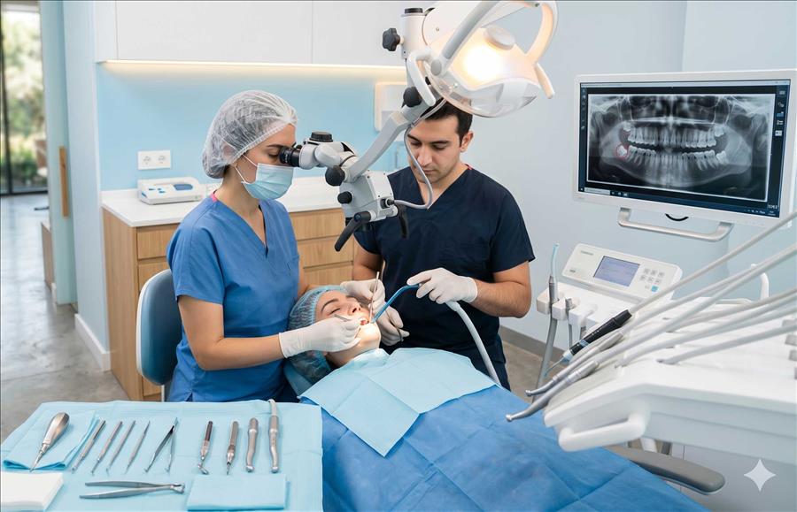
The primary etiology of tooth impaction stems from evolutionary adjustments leading to a gradual reduction in human jaw size. When fully formed permanent teeth lack the required arch perimeter to erupt, they become trapped behind a dense barrier of bone or fibrous tissue. In cases of partial impaction, a segment of the crown breaks through the gingiva while the remainder of the clinical crown and root structure stays locked beneath the surface. Before scheduling impacted tooth operations, a comprehensive radiological evaluation is paramount to establish an accurate clinical roadmap. Utilizing panoramic X-rays and, when necessary, advanced three-dimensional digital cone-beam computed tomography (CBCT), oral surgeons can map the exact location, root curvature, and precise proximity of the tooth to critical anatomical landmarks such as the inferior alveolar nerve.
Not every impacted tooth requires immediate extraction; however, surgical removal becomes a medical necessity when a tooth shows a clear pathological tendency or compromises surrounding tissues. Key clinical indicators that prompt a definitive recommendation for surgical intervention include:
With modern advancements in minor oral surgery and highly effective local anesthesia protocols, impacted tooth extractions are performed as completely pain-free, comfortable outpatient procedures. At Akçadent Dental Clinic, we maintain the highest medical sterilization standards and prioritize patient comfort through every phase of treatment. During the procedure, a precise micro-incision is made in the gingival tissue to gain safe access to the underlying bone. If the tooth is deeply embedded or locked at an adverse angle, it is carefully sectioned into smaller fragments to preserve the surrounding bone structure during removal. Once the extraction is complete, the site is cleaned thoroughly and closed with biocompatible sutures to promote stable healing.
The long-term success of the surgical procedure depends heavily on the patient’s adherence to post-operative care instructions during the critical initial healing phase. Applying an external cold compress intermittently during the first twenty-four hours significantly reduces post-surgical edema and swelling. For the first few days following surgery, patients must completely avoid hard, crunchy, extremely hot, or highly acidic foods, choosing soft, lukewarm, or pureed options instead. Furthermore, habits that create negative intraoral pressure—such as smoking or drinking through a straw—must be strictly avoided to prevent dislodging the protective blood clot, which can lead to a painful recovery complication known as dry socket (alveolitis). When prescribed antibiotics, analgesics, and antiseptic mouth rinses are used precisely as directed, the surgical site recovers rapidly and smoothly.
Asymptomatic teeth completely embedded in the bone that show no signs of infection, cysts, or damage to adjacent roots can often be monitored through regular dental X-rays. However, partial impactions or teeth showing pathological trends require extraction as a clinical necessity.
The roots of lower impacted teeth often lie in close proximity to the inferior alveolar nerve, which supplies sensation to the lower lip and chin. A preoperative three-dimensional cone-beam computed tomography (CBCT) scan is used to precisely evaluate this anatomy and minimize risk during surgery.
Biocompatible sutures are placed during minor oral surgery to close the surgical site and ensure proper initial clot formation. Depending on the rate of tissue healing, these stitches are gently removed by an oral surgeon during a follow-up appointment, usually 7 to 10 days after the procedure.
Protecting the blood clot that forms in the extraction socket is critical for healthy tissue regeneration. Patients must strictly avoid smoking, spitting forcefully, or drinking through a straw for the first few days, as the resulting negative intraoral pressure can dislodge the clot and cause a painful dry socket.